Listeria Swimming in a Host Fibroblast
Listeria Swimming in a Host Fibroblast
Submitted by Michele Balsamo of the Gertler Lab at the Koch Institute
MIT Department of Biology, Koch Institute at MIT
Michele Balsamo
Gertler Lab, Koch Institute
Deconvolution Micrograph
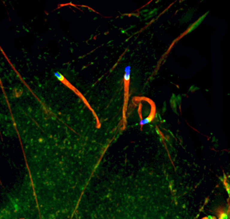
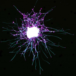
"This image captures three bacteria (Listeria monocytogenes) swimming in the cytoplasm of a host cell (a fibroblast). While outside a cell, Listeria does not move by an actin tail (in red), but by flagella. In infected cells Listeria hijacks the cell motility machinery of the host cell to move in the cytoplasm of the cell and to spread cell to cell. Mena is labeled in green, and we can see that is recruited at the interphase between the bacterium (blue is the bacterial DNA) and the F-actin tail (red). Cell biologists often use Listeria as a model for understanding the basics of actin assembly and cell motility.
This picture was taken quite early during my project. I wanted to understand the contribution of different version of the Mena protein during actin remodeling. One assay for looking at actin polymerization in the cell consists of genetically engineering the host cell with genes supposedly important for actin remodeling and infecting them with Listeria monocytogenes, for looking at the F-actin tail length. Using this assay I found that different Mena versions differently affect the Listeria F-actin tail, and thus actin polymerization."