Localization of Brd4-Nut Fusion in a Cell, Version #1
Localization of Brd4-Nut Fusion in a Cell, Version #1
Submitted by Scott Floyd of the Yaffe Lab at the Koch Institute
MIT Department of Biology, MIT Department of Biological Engineering, Koch Institute at MIT
Scott Floyd
Yaffe Lab, Koch Institute
Deconvolution Micrograph
"Nut-midline carcinoma is a rare but nearly universally fatal cancer that affects mostly young people (children and adults <35 years old). The cancer is defined by the t(15:19) chromosomal translocation that fuses the Nut1 gene to the N-terminus of the Brd4 gene, which is a bromodomain containing protein that interacts with acetylated lysine on histones. Although survival in this cancer is poor, radiotherapy and chemotherapy are the only known effective treatments. Data from our lab indicates that Brd4 can suppress signaling after DNA damage caused by irradiation and chemotherapy, sensitizing the cell to these treatments.
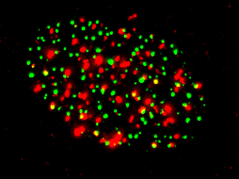
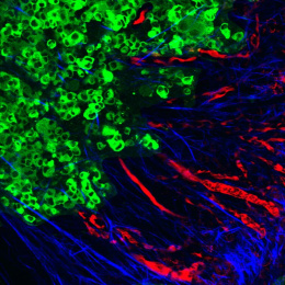
This image shows an irradiated cell and indicates the cellular localization of the product Brd4-Nut fusion protein in green and the DNA damage marker gH2AX in red, indicating that these two markers are mutually exclusive.
We wanted to understand where the Brd4-Nut fusion protein is localized in the cell in relation to both markers of DNA damage signaling and acetylation marks on histones in the chromatin. This gives us insight into the function of this cancer-related molecule to prevent DNA damage signaling, and gives information on how the molecule acts."