Visualizing Inhibitory Neurons with Immunofluorescence
Visualizing Inhibitory Neurons with Immunofluorescence
Submitted by Eitan Kaplan of the Bear Lab at the Picower Institute for Learning and Memory
Picower Institute for Learning and Memory, MIT Department of Brain and Cognitive Sciences
Eitan Kaplan
Bear Lab, Picower Institute for Learning and Memory
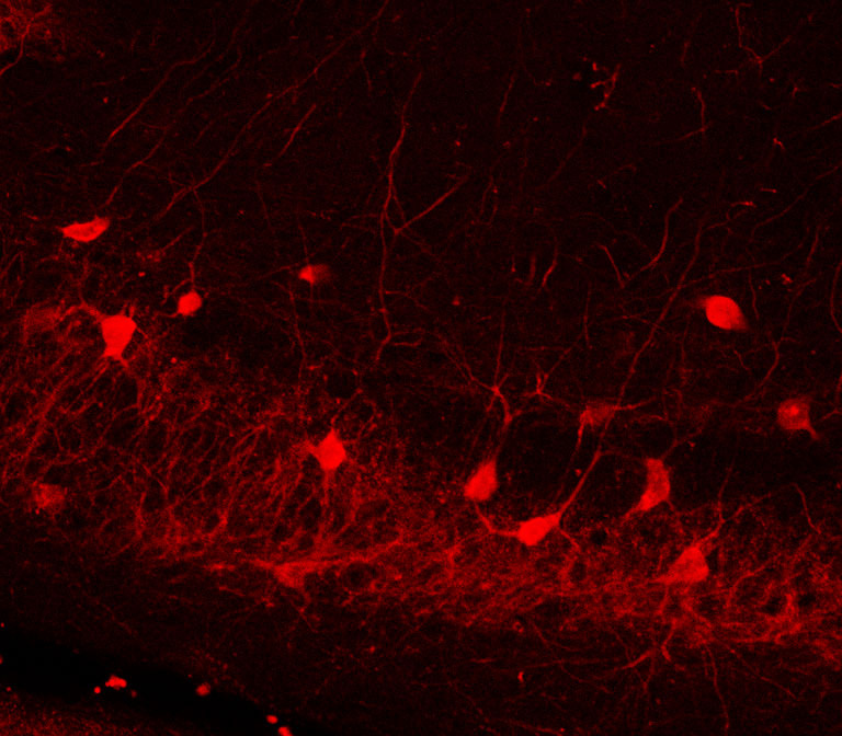
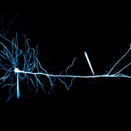
Confocal Micrograph
"In this image we used immunofluorescence imaging to visualize a class of nerve cells called parvalbumin-positive inhibitory neurons. Changes in the number of this neuron type has been implicated in many neurological diseases including schizophrenia. To understand their influence, we need to be able to visualize this specific class of inhibitory neurons."