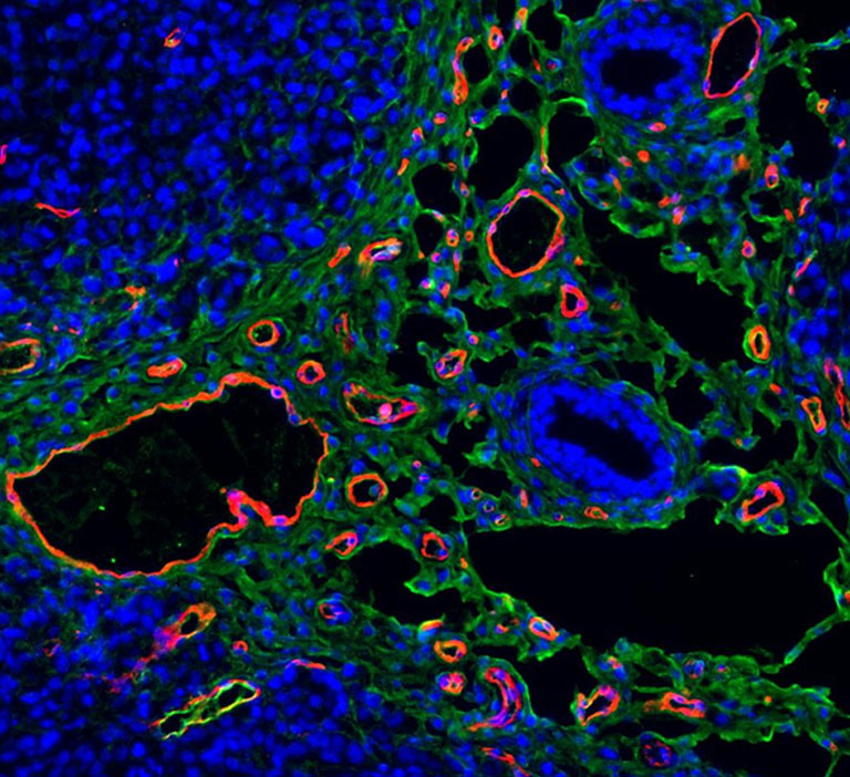
Coagulation Deposits in a Human Tumor
Coagulation Deposits in a Human Tumor
Submitted by Kevin Lin and Sangeeta Bhatia of the Laboratory for Multiscale Regenerative Technologies at MIT
Koch Institute at MIT, Institute of Medical Engineering and Science
Kevin Lin and Sangeeta Bhatia
Laboratory for Multiscale Regenerative Technologies, Koch Institute
Epi-Fluorescence Micrograph
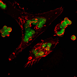
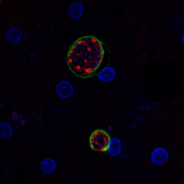
"This image is a cross section of a human tumor grown in an animal host. The stains used in this image are Hoechst (blue) to mark cell nuclei, and immunostaining against CD31 (red) to mark blood vessels, and fibrin (green) to mark coagulation deposits.
The picture was taken to characterize and visualize fibrin deposition in tumor xenografts in response to a relatively new class of chemotherapeutics called vascular disrupting agents (VDAs). We were trying to see if VDAs trigger a significant amount of tumor-specific coagulation activity and whether this activity is confined to vessels or throughout the tumor tissue."