Live Cortical Neurons Labeled with Green Fluorescent Protein
Live Cortical Neurons Labeled with Green Fluorescent Protein
Submitted by Neville Sanjana in the Seung Lab in the Department of Brain and Cognitive Sciences
MIT Department of Brain and Cognitive Sciences, Broad Institute
Neville Sanjana
Seung Lab, Deparment of Brain and Cognitive Sciences
Fluorescence Micrograph
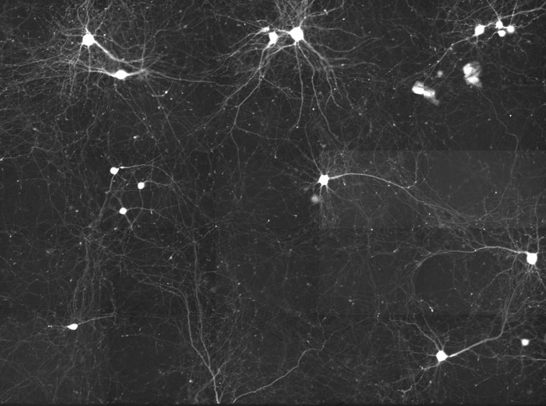
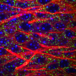
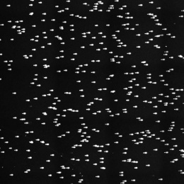
"This image is a still from a time-lapse movie of sparsely GFP-labeled living mouse cortical neurons, imaged on an incubated automated widefield fluorescence microscope over the key period of synaptic development. Axons are the output ends of neurons, transmitting signals from one neuron to multiple downstream neurons in the circuit. I am interested in the trajectories of elongating axons, which perform amazing feats of navigation to wire up a functional brain.
By making time-lapse movies, I (with Sebastian Seung) examined if the growing axons had any stereotyped cellular behaviors and if these behaviors were amenable to quantitative analysis. From the time-lapse movies that contain these stills, we found that each individual axon grows for hours along a remarkably straight trajectory, which is punctuated by infrequent turns. After a turn, the axon again grows straight but in a new direction. How do the axons keep to a straight path? Presently, it is unknown whether axonal straightness is due to an intrinsic property of elongating axons or due to complex signals in the neuron environment."