Metastatic Breast Cancer Invades a Liver, Version #3
Metastatic Breast Cancer Invades a Liver, Version #3
Submitted by Alexandra Naba of the Hynes Laboratory at the Koch Institute
MIT Department of Biology, Koch Institute at MIT
Alexandra Naba
Hynes Laboratory, Koch Institute
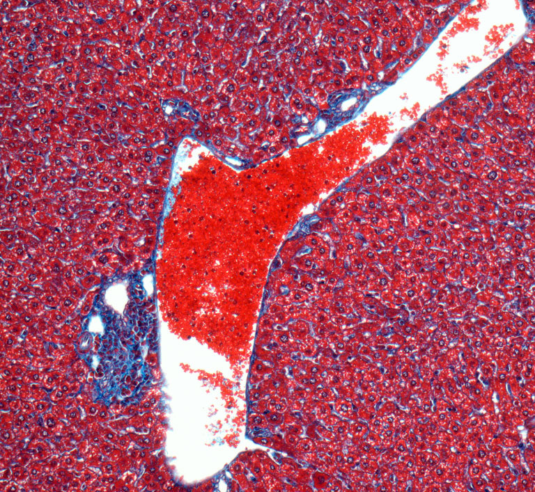
Light Micrograph
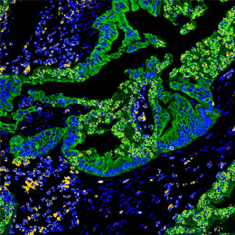
"This image shows a section of the liver of a mouse bearing a metastatic mammary tumor. At the center of the image is a blood vessel that is lined with a layer of collagen fibers called the basement membrane (in blue). One can observe a metastasis coming from the mammary tumor and growing at the very periphery of the blood vessel (collagen-rich structure in the lower left part of the image). The normal hepatic tissue appears in pink/purple (with minimal collagen deposition in blue). The characteristic of the metastasis is the dramatic deposition of collagen fibers (in blue) within and around it. The bright red objects in the lumen of the blood vessel are red blood cells.
I am interested in understanding how the extracellular matrix influences tumor progression and metastasis formation. The extracellular matrix constitutes the architectural scaffold that supports cells within tissues. Alterations in its organization and/or changes in its composition have been shown to promote tumor progression. Collagens – depicted in the images submitted – are one of the main components of the extracellular matrix and I use it as a marker of tumor progression, as the level of extracellular matrix deposition and organization correlates positively with a more advanced stage of tumor progression. My research goal is to characterize the changes in the composition of the extracellular matrix during tumor progression in order to identify novel biomarkers that will serve as prognostic and diagnostic tools for patients with cancer."