Gut Instinct 2: Screening for Signs of Cancer
Gut Instinct 2: Screening for Signs of Cancer
Submitted by Steffen Rickelt of the Hynes Laboratory at the Koch Institute for Integrative Cancer Research
MIT Department of Biology, Koch Institute at MIT
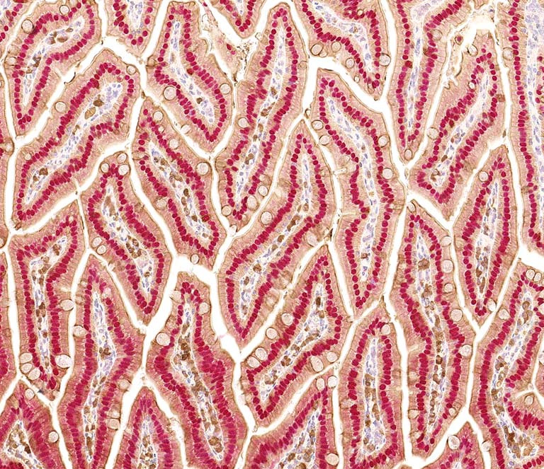
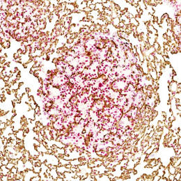
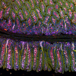
This image shows a cross section through villi of the mouse small intestine. Stained are cell nuclei (red) located to the base of nutrient-absorbing and mucus-secreting columnar epithelial cells (brown) that surround the cellular connective tissue forming the core of each villus.
I primarily took this image for documentation purpose of antibodies against the well-known intestinal marker proteins CDX2 and Pan-Keratin. With this approach I aim to setup multiple immunohistochemical-stainings in our laboratory for their future application on a larger number of human colorectal cancer patient samples.