That's So Fly: Cell Development Pop Art
That's So Fly: Cell Development Pop Art
Submitted by Claudia Vásquez of the Martin Laboratory
MIT Department of Biology
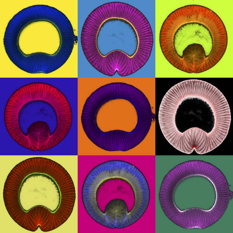
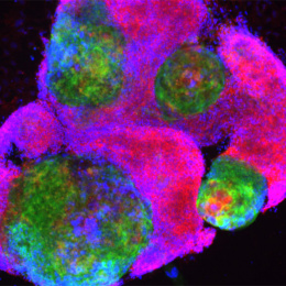
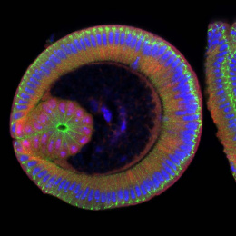
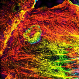
These images are slices from 3 different fly embryos during their early development. It shows how cells coordinate and change shape to ultimately generate a new cell layer.
I study how myosin is regulated during tissue morphogenesis. In our lab we used ventral furrow formation in the fruit fly as a model for tissue morphogenesis. During this process a section of ~1000 cells on the ventral side of the embryo (down) constrict apically (outer perimeter). I use this method to see if the myosin mutants I study localize to appropriate locations within cell and along the whole cell layer. I think this is an example of not only how easily science can be made art but also of how studying something that seemingly as esoteric as fruit fly development is extremely relevant to advancing scientific studies.