Getting Into C. elegans' Head 1
Getting Into C. elegans' Head 1
Rita Droste, Nikhil Bhatla
MIT Department of Biology, McGovern Institute for Brain Research, Koch Institute at MIT
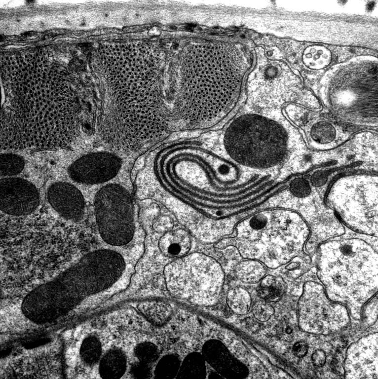
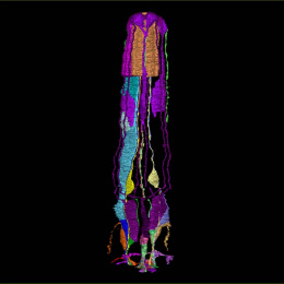
This is an image of a slice of the roundworm C. elegans' head, taken using an electron microscope. In the center of the image, you can see a stacked membrane that happens to look like a serpent. The grid of dark spots in the upper left corner is a set of muscle filaments, and the large dark blobs throughout the image are mitochondria, the energy-producing organs of the cell. Several neural fibers are also visible.
This image was taken in the context of a serial section reconstruction of the worm's feeding organ, the pharynx. We were studying the synaptic connectivity between neurons and muscle visible only by electron microscopy.