The Whole Whorl in its Pharynx
The Whole Whorl in its Pharynx
Rita Droste, Nikhil Bhatla
MIT Department of Biology, McGovern Institute for Brain Research, Koch Institute at MIT
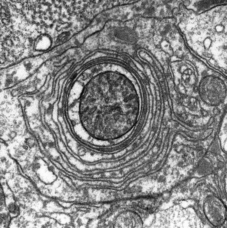
This is an image of a slice of the roundworm C. elegans' head, taken using an electron microscope. In the center of the image, you can see a large whorl of membrane found within a sheath cell. The exact function of this organelle is unknown.
This image was taken in the context of a serial section reconstruction of the worm's feeding organ, the pharynx. We were studying the synaptic connectivity between neurons and muscle visible only by electron microscopy.
We are interested in how different cells in the worm interact, and electron microscopy analysis helps develop hypotheses for how a neuron might synapse and therefore communicate with another cell.