Confirming How Worm Sperm Form
Confirming How Worm Sperm Form
Rita Droste, Christoph Engert
MIT Department of Biology, McGovern Institute for Brain Research, Koch Institute at MIT
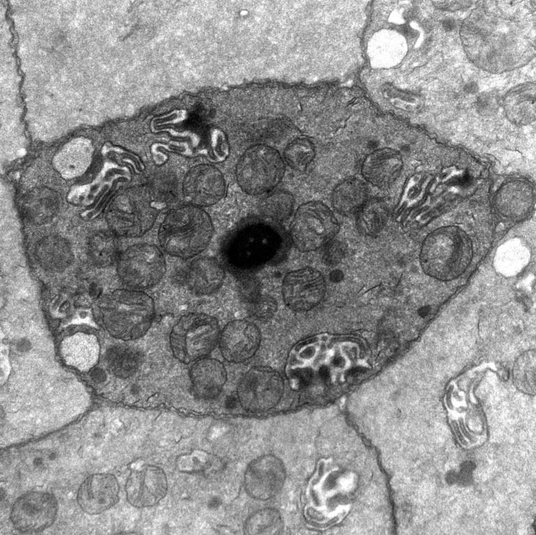
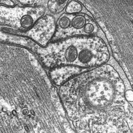
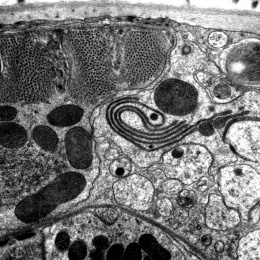
This is an image of a slice of the roundworm C. elegans' reproductive tract, taken using an electron microscope. In the center of the image, you can see a worm sperm, with a dark, compacted nucleus.The round, grey structures are mitochondria, which provide the energy needed for fertilization. The tentacled, white objects are seminal vesicles, which comprise a head, collar and folded membranes. They activate sperm motility so that the sperm can crawl to the egg to fertilize it.
This image was taken to study the shape of sperm. We have found mutant worms that have fewer sperm, and we were curious about whether the sperm developed normally. Specifically, we were interested in the compaction of the nucleus which is a critical step to prepare the sperm genome for fertilization. This image shows a normal worm sperm.