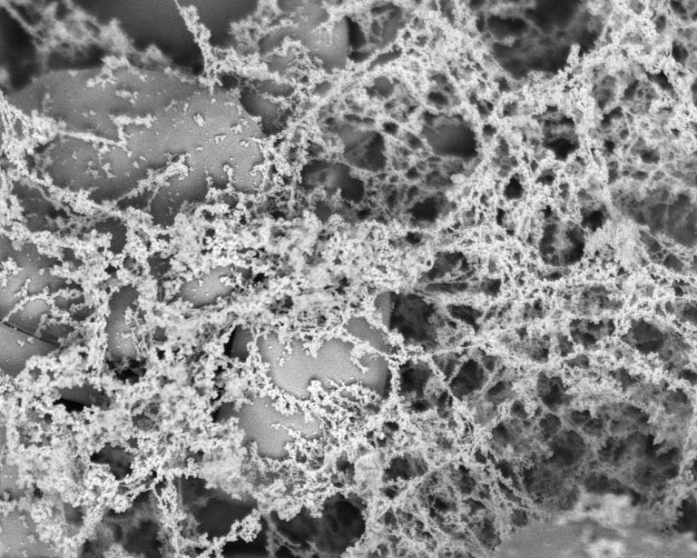
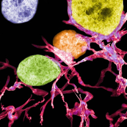
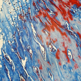
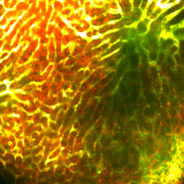
Blood Clots with Nanofiber 2
Blood Clots with Nanofiber 2
Bryan B. Hsu, Shuguang Zhang and Paula T. Hammond, Koch Institute at MIT
Koch Institute at MIT, MIT Department of Chemical Engineering
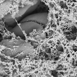
This image shows nanofiber (RADA16-I) clots with blood cells. The images in this series show what a blood clot looks like on a microscopic scale. You can see the blood cells mixed in with either fibrin (the naturally occurring protein that assembles to give mechanical strength to a blood clot) or a short peptide (RADA16-I) that spontaneously self-assembles to entrap the blood cells, forming an artificial clot. By taking these images, we wanted to learn how RADA16-I interacted with blood and we found that they appear to do so in a similar fashion to fibrin.