Nanoparticles in an Adhesive Hydrogel
Nanoparticles in an Adhesive Hydrogel
Credit: Nuria Oliva Jorge
Koch Institute at MIT, Institute of Medical Engineering and Science
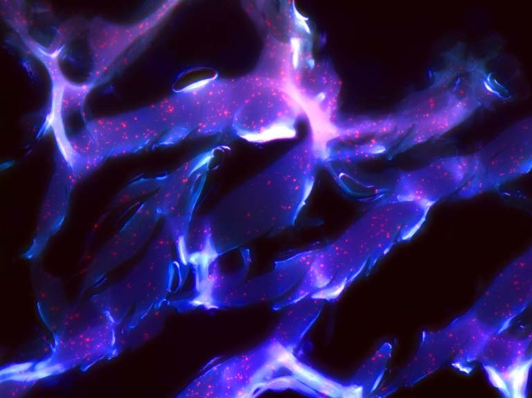
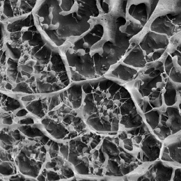
This image shows the porous microstructure of an adhesive hydrogel (blue) that acts as a scaffold for cancer-combating nanoparticles (red dots). Once the adhesive hydrogel is applied to the tumor site, the nanoparticles are released and attack cancer cells.
I took this image to be able to visualize the three-dimensional structure of the material and the relative position of the nanoparticles with respect to the adhesive. The purpose for this was gaining insight into the material-nanoparticle interactions, which would in turn report on release kinetics, critical for effective cancer treatment.
The image shows that there are interactions between the adhesive and the nanoparticles, as inferred by the overlay of the nanoparticles with the material three-dimensional structure. Given the ionic nature of both components, the interactions are mainly electrostatic. These interactions were used explain the sustained release over a short period of time (approximately two days), as opposed to a release over a few hours that would have been observed if no interactions had occurred.