Eye of the Zebrafish 3
Eye of the Zebrafish 3
Dahlia Perez, Adam Amsterdam, Jacqueline Lees
MIT Department of Biology, Koch Institute at MIT
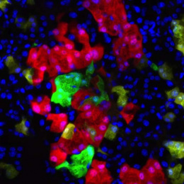
Uveal melanoma (UM) is the most common adult primary eye tumor, and though successful treatments exist for the primary tumor, half of UM patients develop metastases within 15 years at which time their average survival is 6 months. Therefore, a desperate need exists for improved understanding and treatment of this disease. We have generated a model of UM in zebrafish driven by the same genetic changes that are responsible for inducing human UMs, specifically activating mutations in the GNAQ/GNA11 genes. In combination with other cancer-causing mutations, our fish develop melanomas with high frequency and die of this disease.
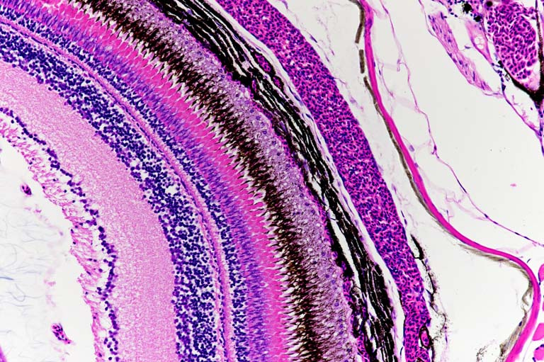
In the picture above, the retina of a GNAQ/11 transgenic zebrafish is brought into focus. Based on the view from this angle of incision, the normal anatomy of the zebrafish eye will begin with the lens at the left side and proceed across the retina in ordered layers of various nerve and photoreceptor cells before finally ending in two pigmented cell layers. It is in the second of these pigmented layers, a vascular layer called the choroid, that the cancer forms in both the human malignancy. Though this zebrafish has not developed a choroidal tumor, the choroid layer is thickened in comparison to wild- type zebrafish. This thickening of the choroid is one of several pigmentation defects in the transgenic zebrafish that indicates how the presence of oncogenic GNAQ/11 is sufficient to disrupt melanocyte biology even in the absence of other cooperating mutations.