Human-Derived Colon Cancer Metastasis in Mouse Liver Tissue 1
Human-Derived Colon Cancer Metastasis in Mouse Liver Tissue 1
Steffen Rickelt and Jatin Roper
MIT Department of Biology, Koch Institute at MIT
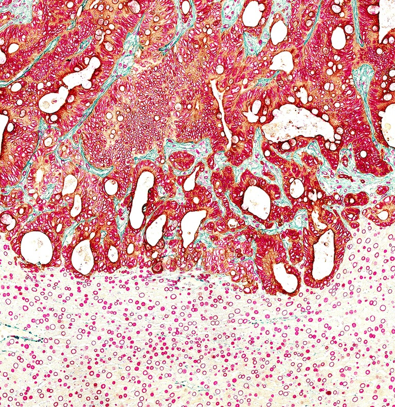
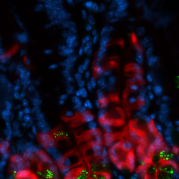
This image shows a section through a mouse liver in which human colorectal cancer-derived organoids (orthotopically-transplanted in the mouse colon) have spread. This liver metastasis shows the invading tumor front (brown tumor cell) - growing from the top of the images towards the bottom - embedded in increasing surrounding connective tissue (green fibers). All nuclei of human tumor cells and the mouse hepatocytes in the lower part of the image were stained with a nuclear membrane marker (red round shapes). Slight green staining in the lower part of the image shows connective tissue that surrounds blood vessels in normal liver tissue.
We took this image to demonstrate metastases formation and thus infiltration and invasion of human-derived, intestinal-specific colon cancer cells (using a human-specific Keratin 20 antibody which is also an intestinal marker) in mouse liver tissue (using a Lamin-A antibody which is a nuclear membrane marker that stains all cell nuclei, human tumor and mouse hepatocytes).
We anticipate to study colon cancer progression in mice to understand mechanisms involved in early tumor cell invasion and metastases with direct relevance for colorectal cancer patients. With our work, we aim to improve early diagnosis and prognosis of colorectal cancer patients in order to expand the lifespan of patients suffering from this disease.