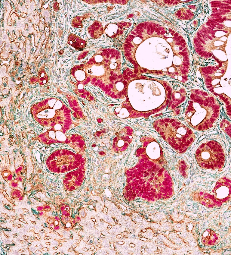
Human-Derived Colon Cancer Metastasis in Mouse Liver Tissue 3
Human-Derived Colon Cancer Metastasis in Mouse Liver Tissue 3
Steffen Rickelt and Jatin Roper
MIT Department of Biology, Koch Institute at MIT
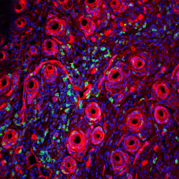
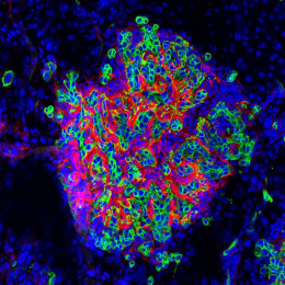
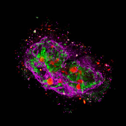
This image shows a section through a mouse liver in which human colorectal cancer-derived organoids (orthotopically-transplanted in the mouse colon) have spread. This liver metastasis shows invading human tumor cell clusters which can be identified by the red nuclear stain. These tumor clusters are surrounded by elongated mesenchymal-derived fibroblast-like cells (green) which are multiplied in the tumor surrounding connective tissue areas. The brown staining shown epithelial-derived cells, i.e. human tumor cells (darker brown) and mouse hepatocytes (lighter brown). We took these images to demonstrate the process of metastases formation and thus infiltration and invasion of human-derived, intestinal-specific colon cancer cells (using an intestine-specific CDX2) in mouse liver tissue.