Mapping Mesenchymal Cells with Vimentin
Mapping Mesenchymal Cells with Vimentin
Marit van Gorsel, Daniel Schmidt, Matthew Vander Heiden
MIT Department of Biology, Koch Institute at MIT
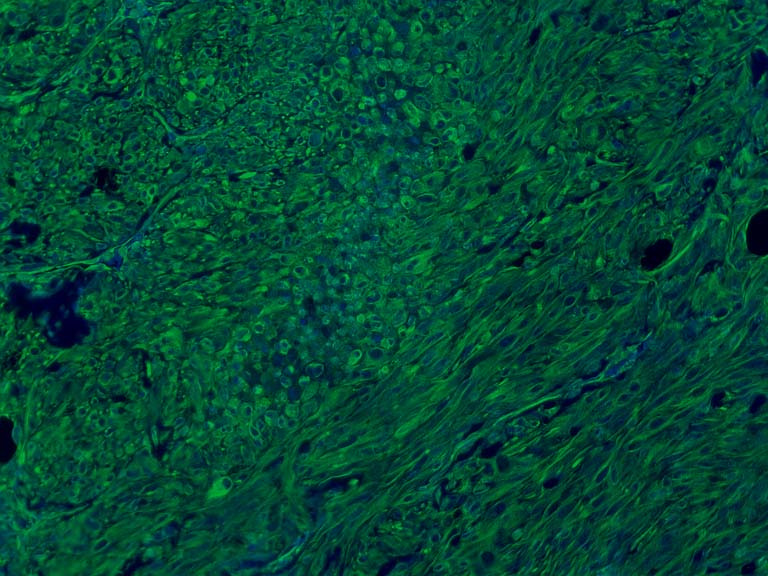
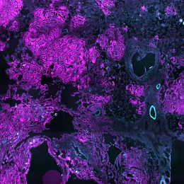
This picture depicts the structural protein vimentin (shown in green) as an example of the organized chaos that exists in a solid tumor. In this case the tumor is a xenograft composed of prostate cancer cells.
This picture was taken to determine whether the cells present in the tumor are more epithelial or mesenchymal. The image shows strong and uniform expression of the structural protein vimentin, which marks mesenchymal cells or cells that have undergone epithelial-mesenchymal transition. This process has been shown in some cases to promote tumor spreading (metastasis).