Behind the Scenes 2
Behind the Scenes 2
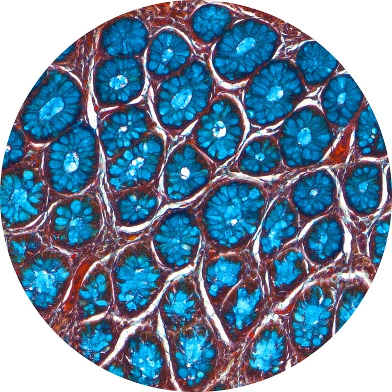
Kaitlyn Sadtler, Corina MacIssac, Robert Langer, Daniel G Anderson
Koch Institute at MIT, MIT Department of Chemical Engineering, Institute of Medical Engineering and Science
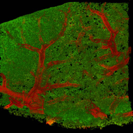
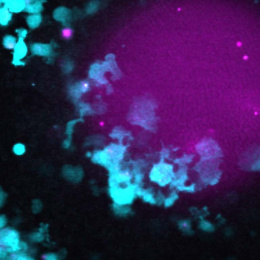
This image, which I like to think of as a “behind the scenes” image, shows one facet of science that is often missed – optimizing and testing the materials you will use in your experiments. Here, we are testing a histochemical stain called Movat’s Pentachrome, which stains the cross-section of a piece of tissue (colon) with 5 different colors of dyes that label 5 large categories of structures found in our tissues: (1) Collagen, Yellow; (2) Nuclei, Dark Purple/Black; (3) Elastic Fibers, Black; (4) Muscle, Red; (5) Mucin, bright blue. Scientists often pick “control” tissues when testing new stains; these are tissues that we know have all of the different structures that we are staining for, so if in our experimental sample, we do not see one color, we can be sure that it was not due to a technical error, and that that structure is not highly abundant in our unknown samples.