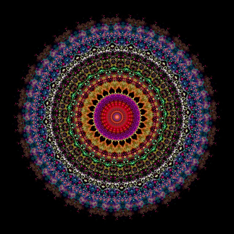
Cellular Mandala: A Microkaleiodoscope of Bioengineering
Cellular Mandala: A Microkaleiodoscope of Bioengineering
Justin Voog, MD, PhD; Neal Peterson, MFA; Sangeeta Bhatia, MD, PhD
Koch Institute at MIT, Institute of Medical Engineering and Science
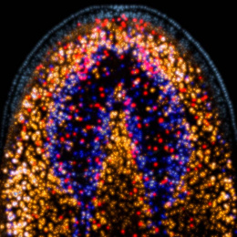
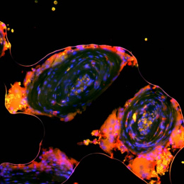
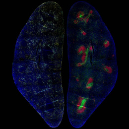
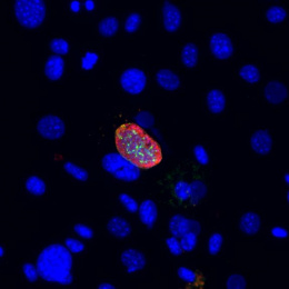
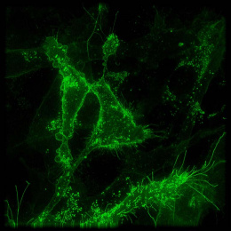
This collage of micrographs highlights the complex and complementary interactions between liver cells (hepatocytes) and supportive stromal cells. Within this image we are able to visualize fluorescently labeled proteins produced by stromal cells that are important for hepatocytes grown in the laboratory. This image is an example of the biologic beauty that can be observed (and created) when science and engineering are coupled.
A major goal of our work is to develop in vitro liver models to better understand regenerative cues that are exploited in liver cancer and metastasis. This set of experiments identified the cellular distribution and expression of stromal factors (and potential therapeutic targets) necessary for maintaining liver models in the lab.