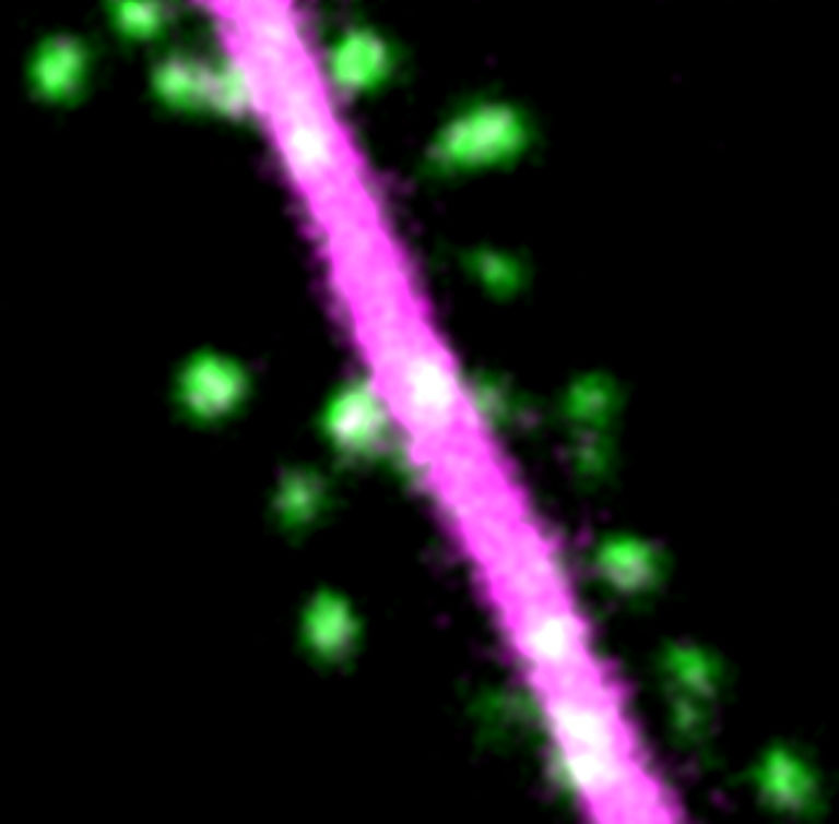
Dendritic Spines in a Hippocampal Neuron
Dendritic Spines in a Hippocampal Neuron
Submitted by Miquel Bosch of the Bear Lab at the Picower Institute for Learning and Memory
Picower Institute for Learning and Memory, MIT Department of Brain and Cognitive Sciences
Miquel Bosch
Bear Lab, Picower Institute for Learning and Memory
Two-Photon Micrograph
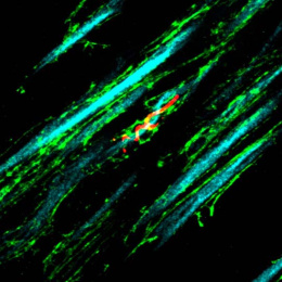
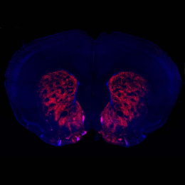
"This is a two-photon microscopy image of a live rat hippocampal neuron transfected with DsRed2 to visualize the dendrite (magenta) and co-transfected with GFP-CaMKII-beta (green) to highlight the structure and dynamics of dendritic spines.
Spines are plastic structures, both physiologically and morphologically. They can become stronger and larger, or weaker and smaller, depending on the input activity. The coordinated plasticity of millions of spines allows the continuous storage of information in neuronal networks. We wanted to visualize in real time if this protein (CaMKII), essential for synaptic plasticity, travelled form one spine to another when one single spine was selectively stimulated. Our larger goal is to unravel the molecular mechanisms of learning and memory at the most fundamental level: the individual synapse."