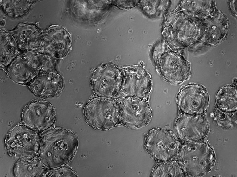
Microtissue Spheres Stick to One Another
Microtissue Spheres Stick to One Another
Submitted by Cheri Li of the Laboratory for Multiscale Regenerative Technologies at the Koch Institute
Koch Institute at MIT, Institute of Medical Engineering and Science
Cheri Li, Sangeeta Bhatia
Laboratory for Multiscale Regenerative Technologies, Koch Institute
Phase Contrast Micrograph
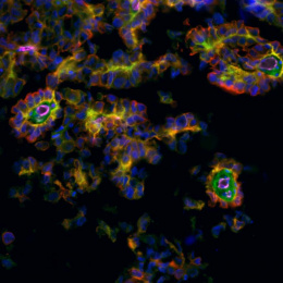
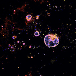
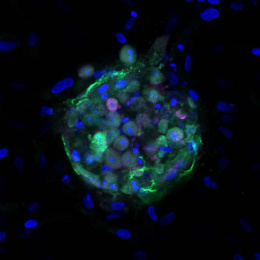
"This image depicts cell-laden hydrogel microtissues aggregated by cell adhesion. Each microtissue (seen in this cross section as a small ball) is a sphere of polymers and hepatocytes. The center aggregate of four small microtissues is held together with additional cells that were added after the microtissue spheres was formed, so that the external cells (seen on the outer edge of the cross-sectioned spheres, and at the points of contact between them) serve as a ‘cellular glue’, and make an aggregate, continuous microtissue of several smaller ones. We’d like to be able to provide a 3D patterned environment to test different combinations of cells, at different densities and organizations, in the hopes of creating micro organs that might be suitable for implantation in a host, for drug and toxicity testing, and perhaps even for living organ support, repair or replacement."