Breaking Rank: How Identical Cells Take on Different Fates
Breaking Rank: How Identical Cells Take on Different Fates
Collections: Image Award Winners
2012 Award Winner
Ni Ji
Van Oudenaarden Lab
Koch Institute at MIT, MIT Department of Biology, MIT Department of Physics
Epi-Fluorescence Micrograph
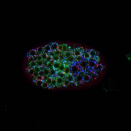
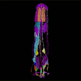
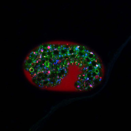
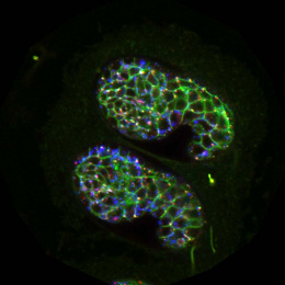
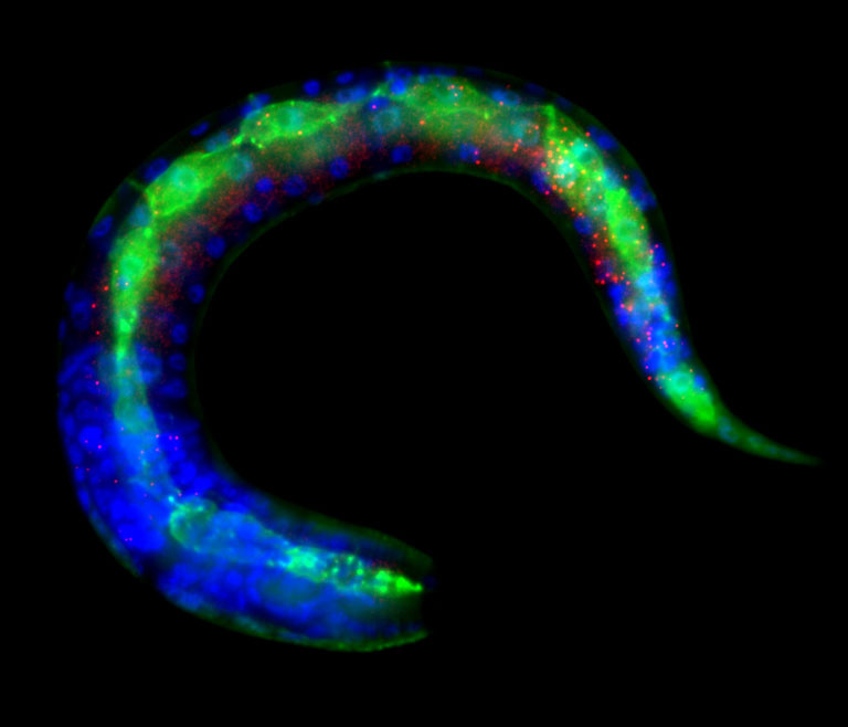
A multicellular organism like a human develops, incredibly, from just one embryonic cell. This image captures a snapshot of this process in a developing worm. At this moment, two seemingly identical neural stem cells move apart and begin to give rise to cells that will be distinct and specialized in the adult worm.
Video
Ni Ji explains how and why she captured this snapshot of development in a worm.