Breast Cancer Cells Spread Over a Textured Surface, Image #3
Breast Cancer Cells Spread Over a Textured Surface, Image #3
Submitted by Julio M. D'Arcy, Erik C. Dreaden, and Paula T. Hammond of the Koch Institute
Koch Institute at MIT, MIT Department of Chemical Engineering
Julio M. D'Arcy, Erik C. Dreaden, Paula T. Hammond
Hammond Laboratory, Koch Institute
Electron Microscopy
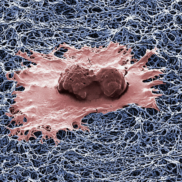
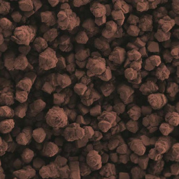
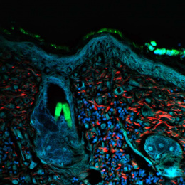
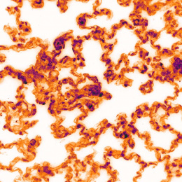
"This image shows a human breast cancer cells (red color) spreading over a porous and texturized polymeric surface (blue color). This cell is supported on nanofibers of an electrically conducting plastic serving as a scaffold for probing cellular growth. These nanofibers can puncture a cell and can be used to inject them with a variety of chemicals that are capable of killing them.
I collected this image in order to study the interaction between a porous polymeric nanofibrillar surface and human cancer breast cells. I was trying to study how fast these cells would spread on a highly doped conducting polymer surface such as 3,4-polyethylenedioxythiophene (PEDOT)."