Nerve Cell Axons Avoid the Slit
Nerve Cell Axons Avoid the Slit
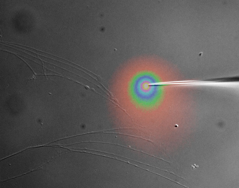
This image, captured by Russell McConnell at the Koch Institute, shows a time-lapse sequence of two nerve cell axons.
MIT Department of Biology, Koch Institute at MIT
Russell McConnell
Gertler Laboratory, Koch Institute
Differential Interference Contrast and Wide-Field Fluorescence Microscopy
"Pictured is a time-lapse sequence of two nerve cell axons. The axons start out on the left side of the image and move to the right to avoid a gradient of a protein called Slit that has been applied with a needle (pseudo-colored to show concentration). We are attempting to understand how the Slit signaling pathway uses the Ena/VASP family of proteins to control cell movement. During development, these spinal neurons send long processes up to the brain to relay information from the peripheral to central nervous system. These processes navigate using proteins found on the surface of other cells as chemical guideposts. In the case of spinal neurons, the Slit protein acts as a Wrong Way sign and redirects the axons."