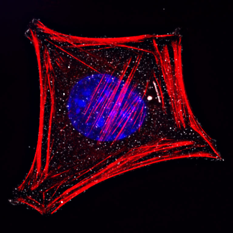
Cancer Cells Create Invadopodia
Cancer Cells Create Invadopodia
Submitted by Tsukasa Shibue in the Weinberg Laboratory at the Whitehead Institute
MIT Department of Biology, Whitehead Institute, Koch Institute at MIT
Tsukasa Shibue
Weinberg Laboratory, Whitehead Institute
Deconvolution Microscopy
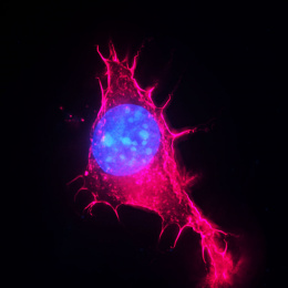
"I am studying various types of cytoskeletal protrusions formed by the cancer cells. Aggressive mouse mammary carcinoma cells (D2A1) were grown on top of a thin gelatin film and stained for the actin cytoskeleton (filamentous actin [F-actin]; red), an actin-binding protein cortactin (white) and the nucleus (by DAPI; blue). These cells developed spots containing F-actin and cortactin, which represent sites where these cells degrade the underlying layer of gelatin film. I aimed to figure out the gelatin degrading/invading ability of the D2A1 cells. This image demonstrated that the D2A1 cells can form actin-rich, cortactin-positive protsuions that invade into the layer of gelatin, which are usually called as ‘invadopodia’.