Epithelial Stain to Track Aneuploidy 2
Epithelial Stain to Track Aneuploidy 2
Submitted by Daniel Eichberg in the Amon Lab at the Koch Institute
Koch Institute at MIT, MIT Department of Biology
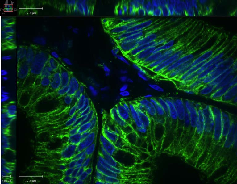
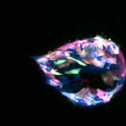
This is a cross section of a surgically removed human colon polyp. The green fluorescent stain is targeting Epithelial Cell Adhesion Molecule (EpCAM), a marker specific to epithelial cells. The blue fluorescent stain is Hoechst, a stain that marks the DNA of all cell types. I took this image to confirm that my EpCAM antibody is able to exclusively target epithelial cells, and not other cell types. Because only the epithelial lining of the polyp has been stained green, and not the fibrovascular core which contains different cell types, I confirmed that my EpCAM antibody exclusively targeted epithelial cells.
Having an abnormal number of chromosomes, a condition called aneuploidy, is a hallmark of cancer cells. Whether aneuploidy causes cancer or is a cause of cancer, and when aneuploidy develops during the progression from normal tissue to late stage cancer, has not been well characterized. To answer these questions, I want to perform single cell sequencing on human tissue samples at various stages of cancer, and see at which stage aneuploidy develops. I will use a cell sorting machine that is able to detect which cells have been labeled with the antibody and emit green light. The cell sorting machine will thus separate epithelial cells that are stained green, from non-epithelial cells that are not stained green, enabling me to sequence only epithelial cells.