Using Sea Urchins to Understand Cell Migration 4
Using Sea Urchins to Understand Cell Migration 4
Genevieve Abbruzzese
MIT Department of Biology, Koch Institute at MIT
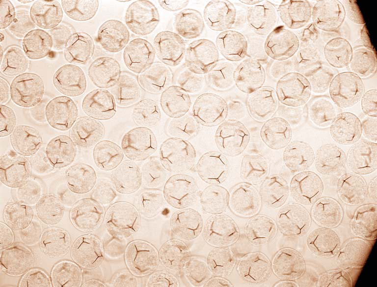
The ability of cells to migrate to distant and precise locations is critical to proper embryo development (as well as cancer metastasis), however there is still much to learn about mechanisms that control this migration and how they find their proper target sites. During sea urchin embryogenesis, when the embryo is still a hollow sphere, mesenchyme cells walk along the inner wall of the embryo until they reach their target site and begin forming the embryonic skeleton. The membrane that makes up the inner wall of the embryo is largely made up of extracellular matrix (ECM), a collection of fibrous proteins and carbohydrates, though it is unknown what proteins are present in this ECM and how they guide and contribute to mesenchyme migration.
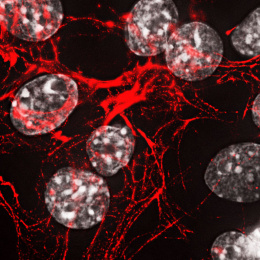
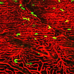
This image shows how we use sea urchin embryos to better understand how this movement happens. We treat the embryos to remove all or most of the cells, leaving behind the fibrous membranes that provide the embryo with organization and structural support. These membranes also serve as the landscape for cells to travel along as they reposition themselves during stages of morphogenesis. Analyzing the protein components that make up this membrane will help us to understand how it acts as a roadmap for motile cells and the signals it retains to guide cells toward their new locations.