Functional Connectivity of Neurons in Three Dimensions
Functional Connectivity of Neurons in Three Dimensions
Murat Yildirim
Picower Institute for Learning and Memory, MIT Department of Brain and Cognitive Sciences
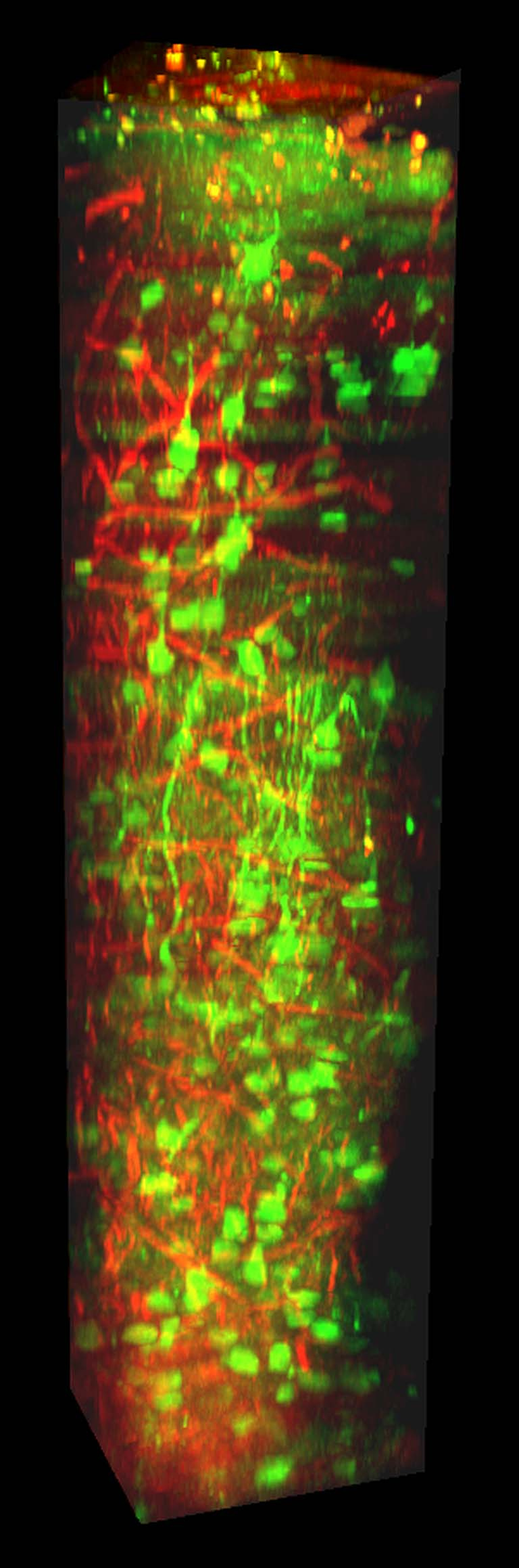
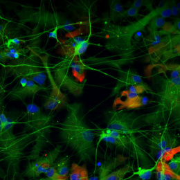
In mammalian brain, visual information acquired from the environment is processed in the primary visual cortex (V1). V1 has six layers and each of these layers have different functions in terms of processing the visual information. In most of the mammal’s brains such as human, primates, and cats, neurons standing in the same layer have similar responses to the same visual stimulus. However, it has been shown that neurons in the superficial layers of the mouse brain have different responses to the same visual stimulus even though they are standing in the same layers. Still, there is a mystery whether neurons in the deep layers of V1 has similar or different responses to the same stimulus. Thus, I developed a home-made microscope which can allow us to image deep layer neurons in V1 of awake mice while we present visual stimulus to the mice. In this image, red color represents blood vessels and green color represent the neurons in all layers of the cortex spanning from superficial (top) to deep layers (bottom) which have similar responses to the same visual stimulus