Investigating Alzheimer's Disease in the Visual Cortex 3
Investigating Alzheimer's Disease in the Visual Cortex 3
Mitchell Murdock
Picower Institute for Learning and Memory, MIT Department of Brain and Cognitive Sciences
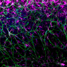
These images are montages of the vasculature of the living mouse visual cortex. I installed a cranial window over the primary visual cortex of a mouse engineered to recapitulate Alzheimer’s pathology.
Each frame depicts a single capillary at different time points. As your eyes pass from left to right and top to bottom, you can see dark patches of cells passing through the capillary. Here, you can see evidence of “stalled” cells, a consequence of severe inflammation in the Alzheimer’s brain. For example, if you look at the frames on the third to last row from the bottom, you can see cells that are stalled, which are reducing perfusion in that brain region. These stalled capillaries are critical factors in reduced blood flow in Alzheimer’s Disease.