Biodistribution of Nanoparticles in a Lymph Node, Warhol Style
Biodistribution of Nanoparticles in a Lymph Node, Warhol Style
Alex Schudel (Postdoctoral Associate, Langer Lab)
Koch Institute at MIT, MIT Department of Chemical Engineering
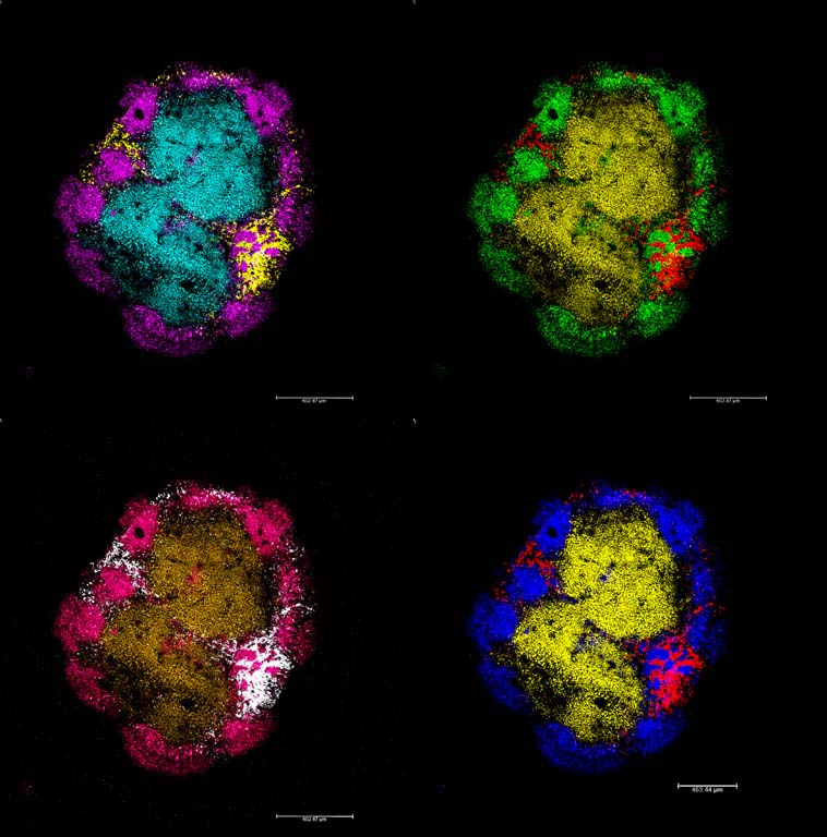
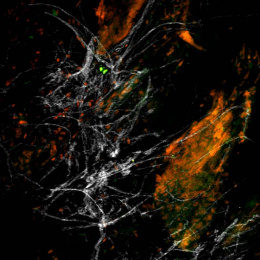
This is an image of a mouse lymph node. It has been stained to target the main cell populations of the adaptive immune system in order to see where nanoparticles that were injected have gone. In the upper left image, the cyan color is the T cells, the purple color is the B cells, and the yellow color is the nanoparticles. This image shows nanoparticle penetration of the lymph node structures and cell locations from the periphery inwards.
We were trying to understand where the injected nanoparticles end up in the lymph node substructure areas in order to determine which cells they might encounter. This technique gives both spatial and cellular understanding about the biodistribution of these nanoparticles within this tissue.