Axons from the Retina Project into the Visual Thalamus, Version #2
Axons from the Retina Project into the Visual Thalamus, Version #2
Submitted by Jason Coleman of the Bear Lab in the Picower Institute for Learning and Memory
Picower Institute for Learning and Memory, MIT Department of Brain and Cognitive Sciences
Jason Coleman
Bear Lab, Picower Institute for Learning and Memory
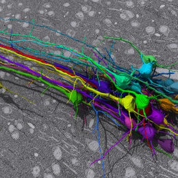
Laser-Scanning Confocal Micrograph
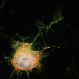
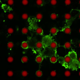
"This image shows axons from the retina of a mouse projecting into the visual thalamus. Different colors correspond to portions of axon branches in different depths of a 3D volume. We took this image to look at the pattern of axon projections from part of one retina to visual thalamus in a volume. The purpose was to illuminate the detailed morphology of axon branches with the hope that it might reveal something about how they function in the visual pathway."