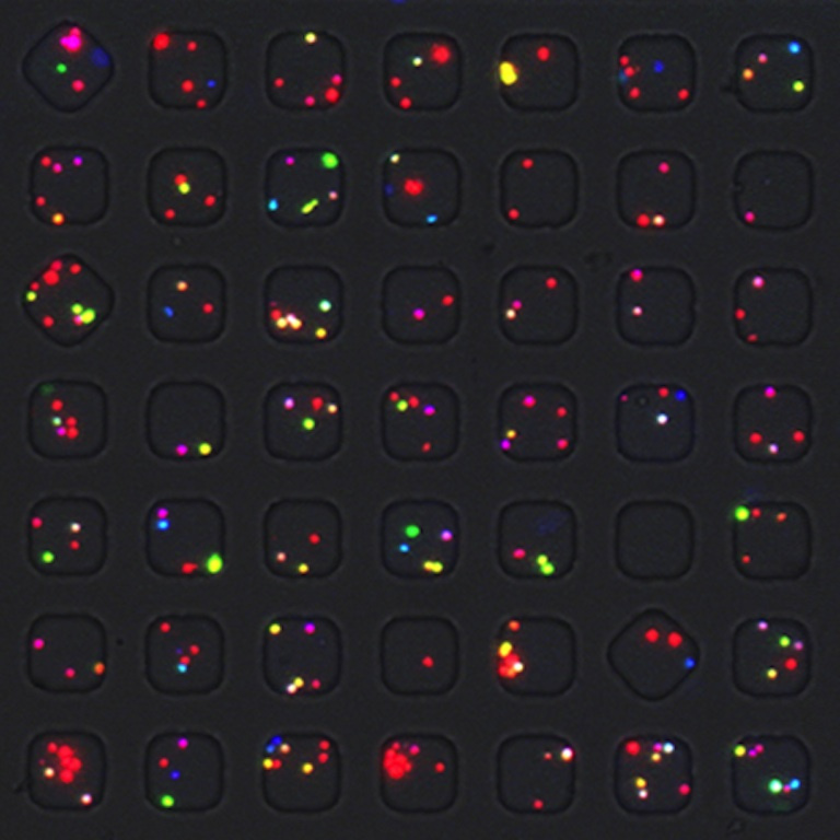
Barcodes For Immune Cells
Barcodes For Immune Cells
Collections: Cancer Immunology & Immunotherapy
Submitted by Yvonne J. Yamanaka & Gregory L. Szeto at the Koch Institute
Koch Institute at MIT, MIT Department of Chemical Engineering, MIT Department of Biological Engineering, MIT Department of Materials Science and Engineering
Yvonne J. Yamanaka & Gregory L. Szeto
Love Lab & Irvine Lab, Koch Institute
Epi-fluorescence Microscopy
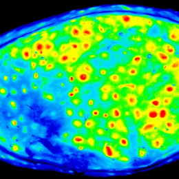
"This image shows human immune cells that are “barcoded” using different combinations of fluorescent dyes. Barcoding allows us to analyze multiple samples of cells at once, which makes experiments more efficient and cost-effective. In this image, the barcode of each cell provides information about what type of stimulation the cell was previously exposed to. The cells are loaded on an array of nanowells. In this image, 49 nanowells are visible; the entire 3” x 1” array contains 84,672 nanowells."