Getting "Ahead" of Zebrafish Development
Getting "Ahead" of Zebrafish Development
Submitted by Isabel Brachmann of the Sive Laboratory at the Whitehead Institute for Biomedical Research
Whitehead Institute, MIT Department of Biology
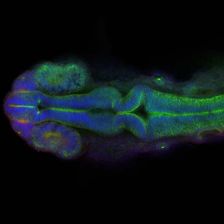
This image shows the brain of a 24 hours post fertilization zebrafish embryo, along with the developing eyes and ears. We study cell shape changes that result in folding of the neural tube, the structure of which brains are formed in the head region. In this image one of the most prominent folds, the midbrain-hindbrain boundary constriction is already formed and the ventricles which will later be filled with cerebrospinal fluid just begin to inflate.