Spatial Expressions: Snapshot of a Growing Tumor
Spatial Expressions: Snapshot of a Growing Tumor
Collections: Image Award Winners, Cancer Discovery Science
2018 Award Winner
Leah Caplan, Jatin Roper, Inbal Avraham-Davidi, Sebastian Santos, Ömer Yilmaz, Aviv Regev
Koch Institute at MIT, Broad Institute, MIT Department of Biology
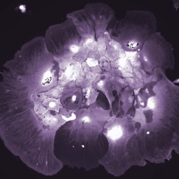
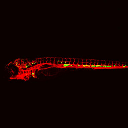
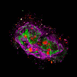
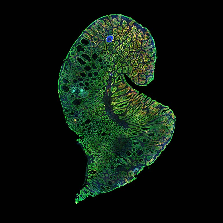
Combining computer vision with single-cell genome sequencing allows researchers to better understand how cells function and interact within the context of their surrounding environment.
In this image, colon cancer cells (green) from a model developed by the Yilmaz Lab have been sequenced and fluorescently marked by the Regev Lab. Yellow tags identify stem-like qualities while red show active proliferation. Together, they present a snapshot of a tumor’s dynamic properties. The team uses this information to determine which biological factors contribute to tumor growth and cancer progression.
This image was taken on a Nikon TI-E with a W1 spinning disk confocal.
Video
Leah Caplan shares the story behind her award-winning image. You can also watch her presentation from the exhibit opening on March 8, 2018 here.