Tumor Cells Travel Through a Fish 1
Tumor Cells Travel Through a Fish 1
David Benjamin
MIT Department of Biology, Koch Institute at MIT
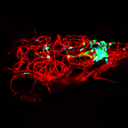
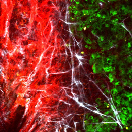
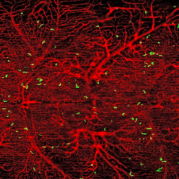
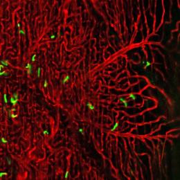
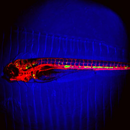
Zebrafish embryos offer a unique system in which to image the events of metastasis. In my experiments, I inject fluorescently-labeled tumor cells into 2 day old zebrafish and then image for periods of hours up to 4 days post-injection. I can watch tumor cells in real time as they travel through circulation, arrest at metastatic sites, exit blood vessels, and form new tumors. The image shows a single 6-day-old fish fish with green tumor cells growing throughout the body 4 days post-injection.