Peripheral Blood Mononuclear Cells Labeled with Barcodes
Peripheral Blood Mononuclear Cells Labeled with Barcodes
Submitted by Yvonne J. Yamanaka of the Love Lab at the Koch Institute
Koch Institute at MIT, MIT Department of Chemical Engineering
Yvonne J. Yamanaka
Love Lab, Koch Institute
Epi-Fluorescence Micrograph
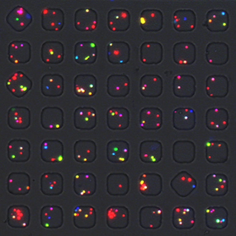
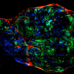
"This image shows human peripheral blood mononuclear cells (PBMCs) that are 'barcoded' by application of different combinations of fluorescent dyes. The barcode of each cell provides information about what type of stimulation condition the cell was exposed to. The cells are loaded on an array of 50 µm x 50 µm x 50 µm microwells. In this image 49 microwells are visible; the entire 3” x 1” array contains 84,672 microwells.
This image allows us to know what types of cells occupy each microwell. Since each cellular barcode color corresponds to a specific stimulation condition, we can build profiles of how immune cells respond to different stimulation conditions by correlating the microwell occupancy information with information about the cytokines and chemokines that are secreted by the cells within each microwell.
In this particular experiment, we used the barcodes to label PBMCs that were stimulated with different doses of a superantigen. We wanted to learn how the dose of superantigen affects the cytokine and chemokine secretion profiles of PBMCs."