Memory Brain Structure Outputs
Memory Brain Structure Outputs
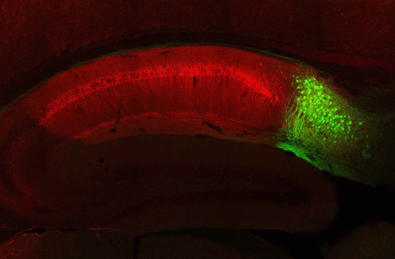
Dheeraj Roy, Susumu Tonegawa
Picower Institute for Learning and Memory, MIT Department of Brain and Cognitive Sciences
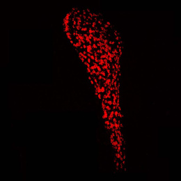
In this image, we are looking at the part of the mammalian (mouse) brain known as the hippocampus, which is conserved in humans. This is where we form new memories and recall past experiences. This memory brain structure has well-known outputs, one is CA1 (shown in red) and the other is subiculum (shown in green). Previous researchers were unable to study subiculum neurons due to their close proximity to CA1. The most exciting part of this image for our research is that by generating a novel transgenic mouse line, we could selectively tag and manipulate subiculum neurons (green) to understand their functional significance. Further, the green subiculum neurons showed no overlap with the red CA1 neurons, which shows the high level of specificity of our novel tool.