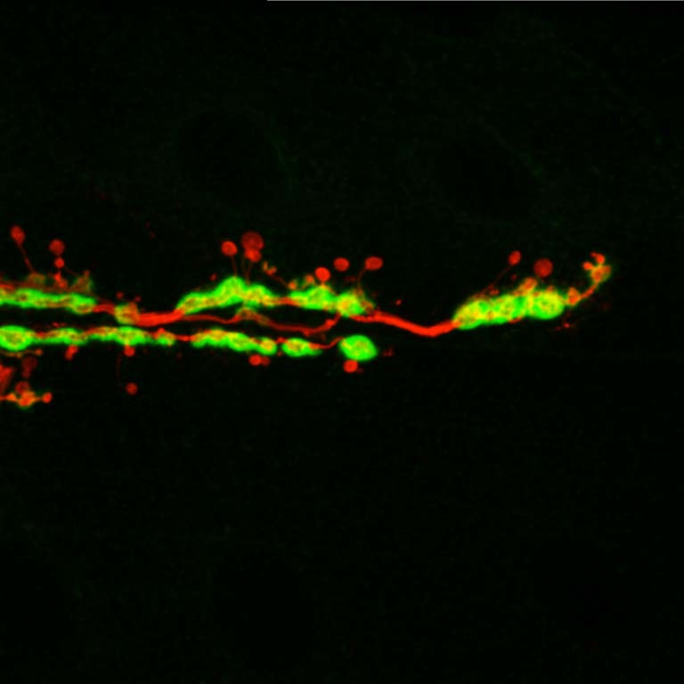
Ghost Boutons and Motor Neurons
Ghost Boutons and Motor Neurons
Submitted by Zachary Piccioli in the Littleton Lab at the Picower Institute for Learning and Memory
MIT Department of Brain and Cognitive Sciences
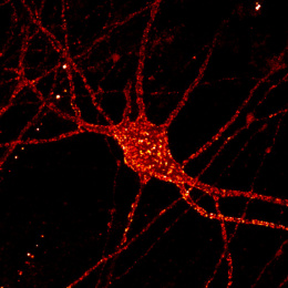
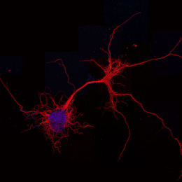
Pictured is a synapse called a neuromuscular junction in the body of a fruit fly larva, where a neuron contacts the muscle it controls. The motorneuron membrane is labeled in red and a postsynaptic scaffold protein within the muscle is labeled in green. This picture shows several neuronal swellings, called boutons, that lack postsynaptic specialization (red without green cloud surrounding). These so-called ghost boutons represent a form of rapid activity-dependent synaptic plasticity that could have implications for the study of learning and memory. We are working to identify the signaling mechanisms that cause ghost bouton budding in response to high activity.